Research Team of Jiří Pacherník
Keywords
myocard, stem cell, cardiomyogenesis, specification of cardiomyocytes
| Head of laboratory: | Mgr. Jiří Pacherník, Ph.D. |  |
| Office: | UKB – A36/111 | |
| E-mail: | jipa@sci.muni.cz | |
| Phone: | 549 49 5578 | |
| Teaching | ||
| Publications | ||
Ongoing projects:
Stem cells are specialized group of cells which are capable of self-renewal and producing same type of cells, but also of differentiating, changing to a different, more specialized type of cell. They play an important role in growth and regulation of homeostasis of animals during their whole development. Study of mechanisms that are responsible for the origin and preservation of stem cells is essential for understanding of basic principles of normal and pathological development of animals.
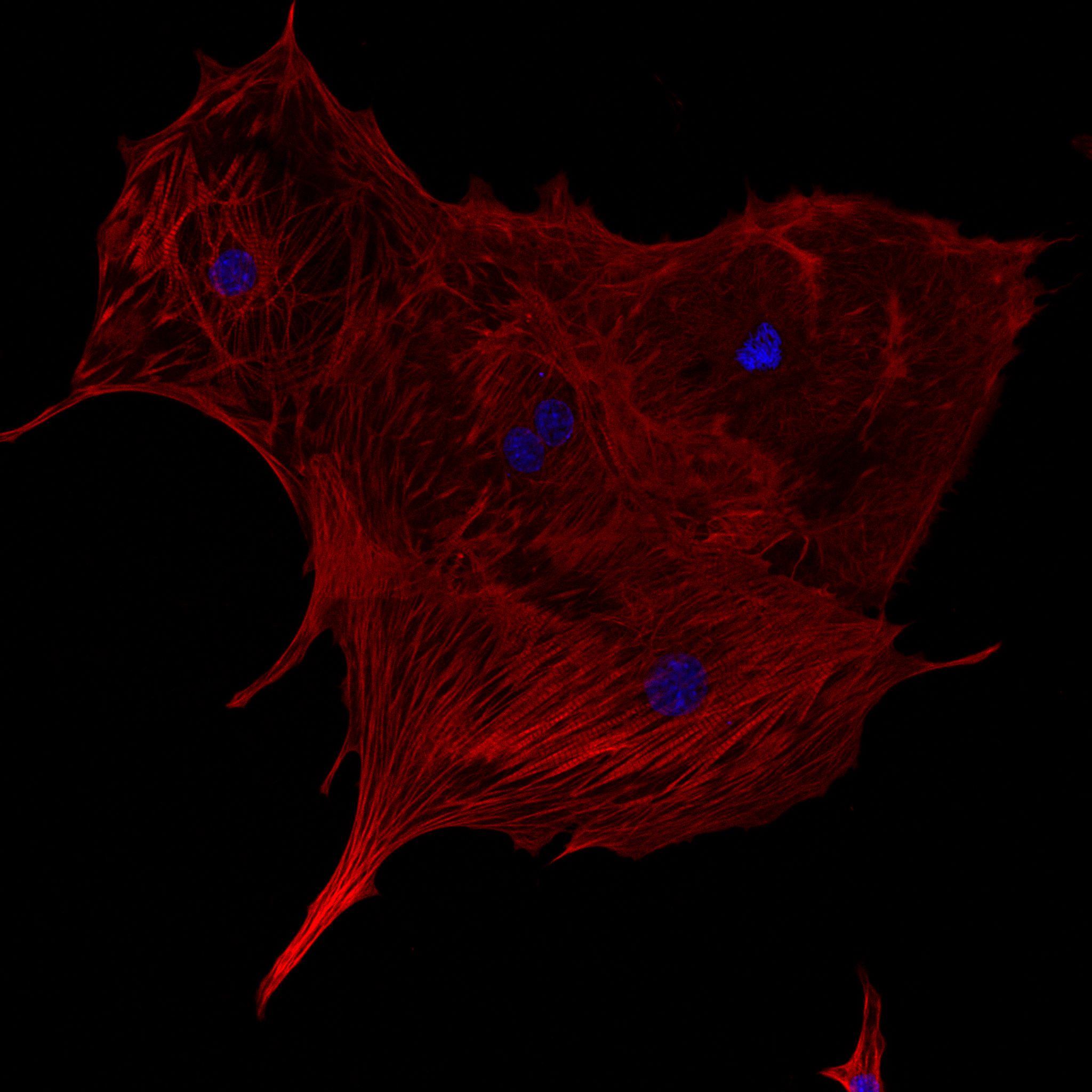
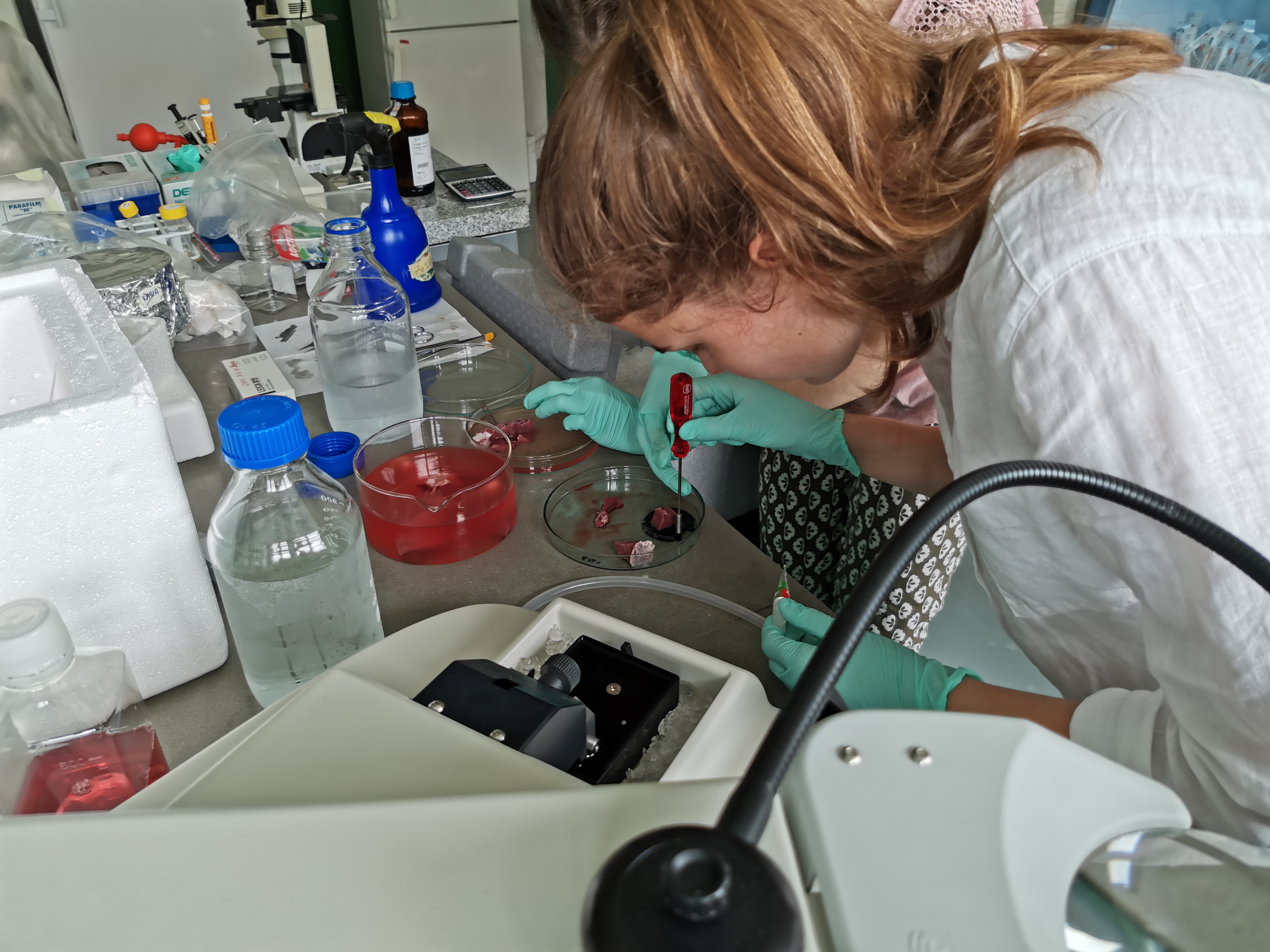
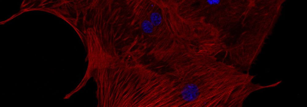
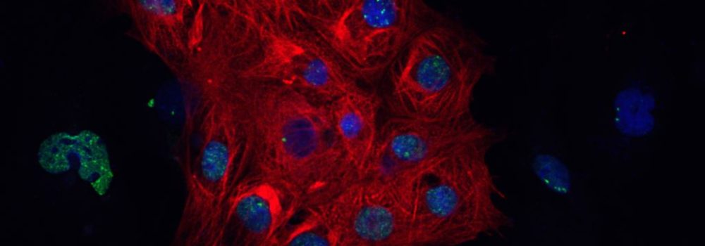
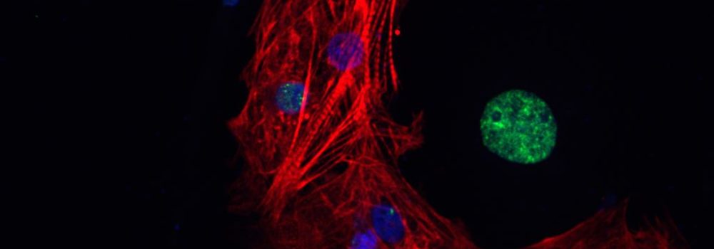
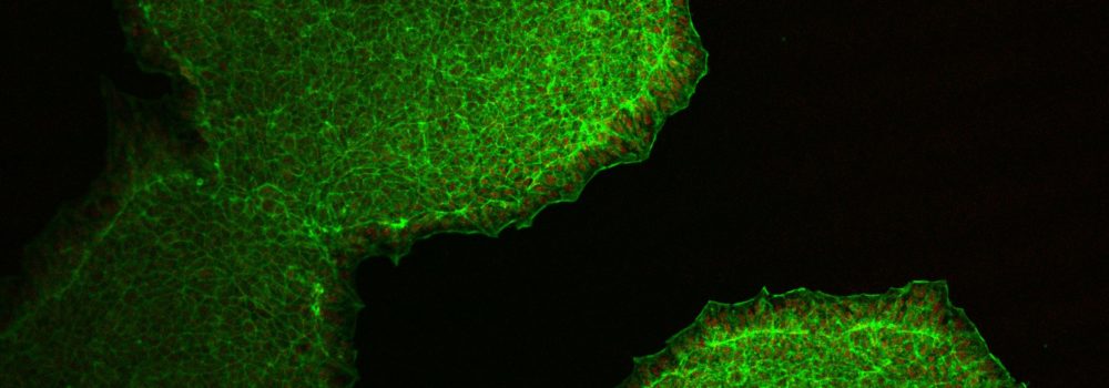
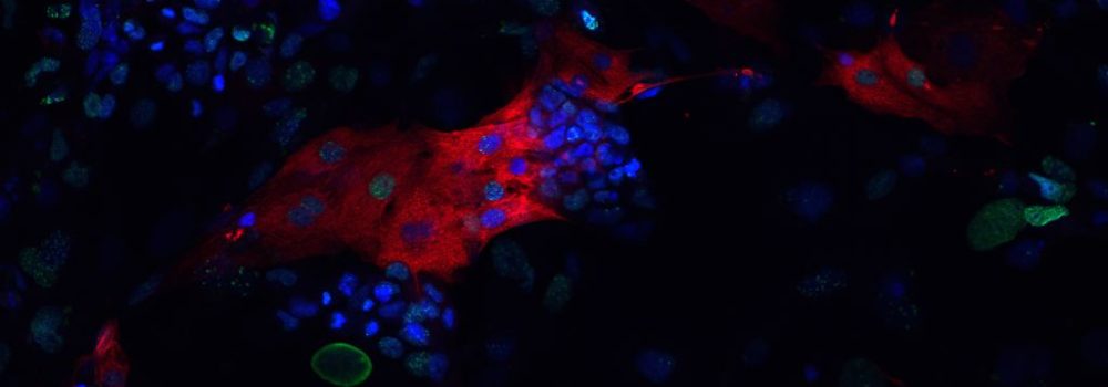
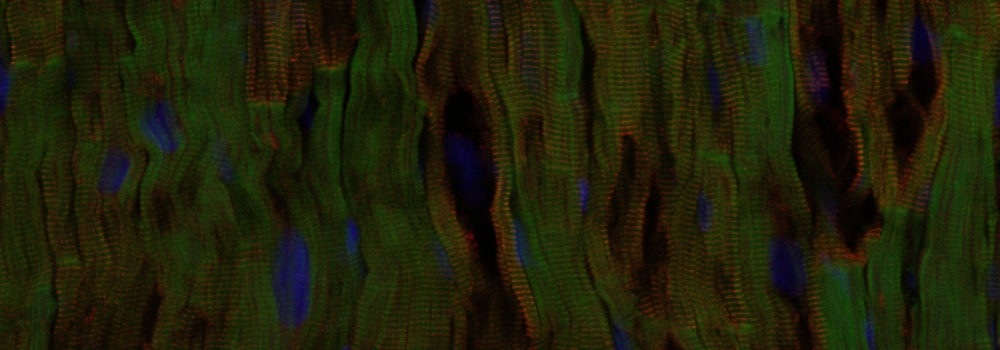
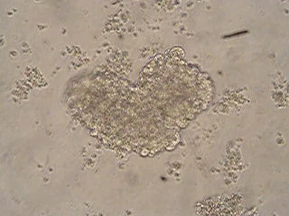
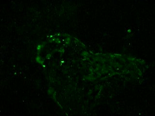
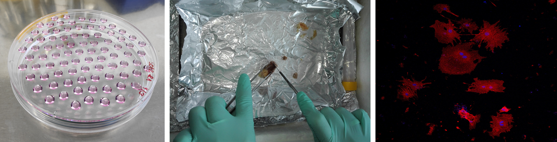
In our laboratory as a model organism we use lab mouse (Mus musculus domesticus) and cell lines that are derived from it as well as some human cell line (mostly human induced pluripotent stem cells). Our experiments are performed on embryonic stem cells and cells that differentiate from them, where we mainly focus on cardiomyocytes.
According to up-to-date research, adult heart does not contain stem cells and its regeneration capacity is very limited. Therefore in case of big damage to myocardium, possible treatments are either whole heart transplantation or transplantation of cardiomyocytes that were prepared from stem cells in vitro. Ideal source for in vitro preparation of cardiomyocytes would be stem cells of myocardium. Cardiac progenitors, which transiently exist during the embryonic development could be considered as model for these type of stem cell. However, in the case of cardiomyocytes that are prepared in vitro, we must also take into consideration what type of cardiomyocytes were actually prepared.
Our research is therefore mainly focused on preparation of myocardial stem cells, preparation and identification of different cardiomyocytes and providing answers to following problems:
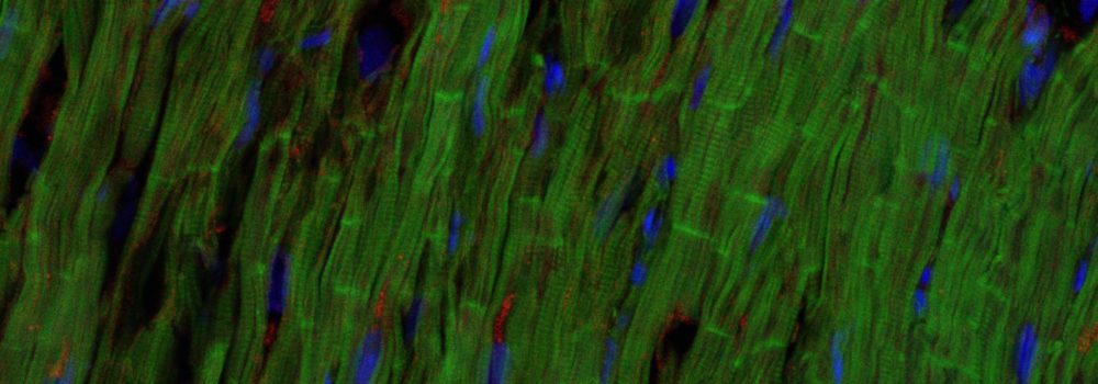
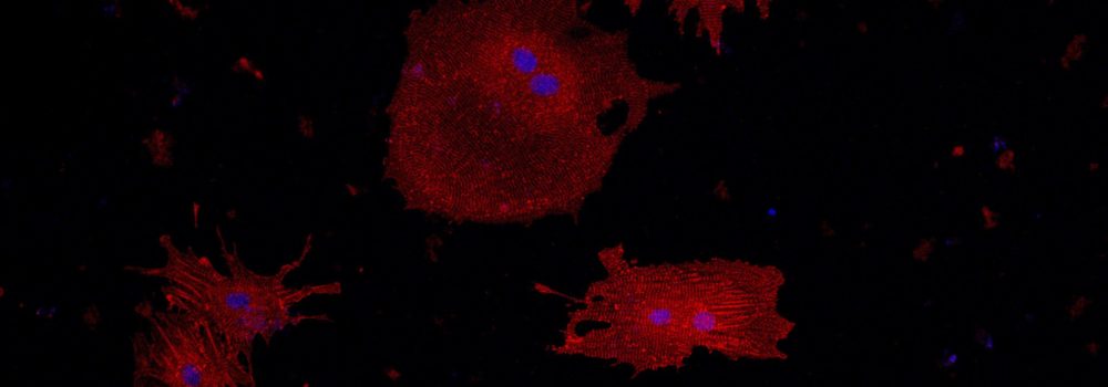
In case of structures that can have some influence on cells in vitro, we are mostly focused on observing changes in characteristics and in differentiation of cells that are cultivated on different kinds of polymers. We are also trying to co-cultivate already isolated cardiomyocytes with other types of cells which can be found in heart (fibroblasts) in 3D structures, where we are studying changes in their morphology and function. When it comes to chemical compounds we are mostly interested in different kinds of inhibitors and activators of various signaling pathways and we are trying to study their effect on differentiation into different types of cardiomyocytes.
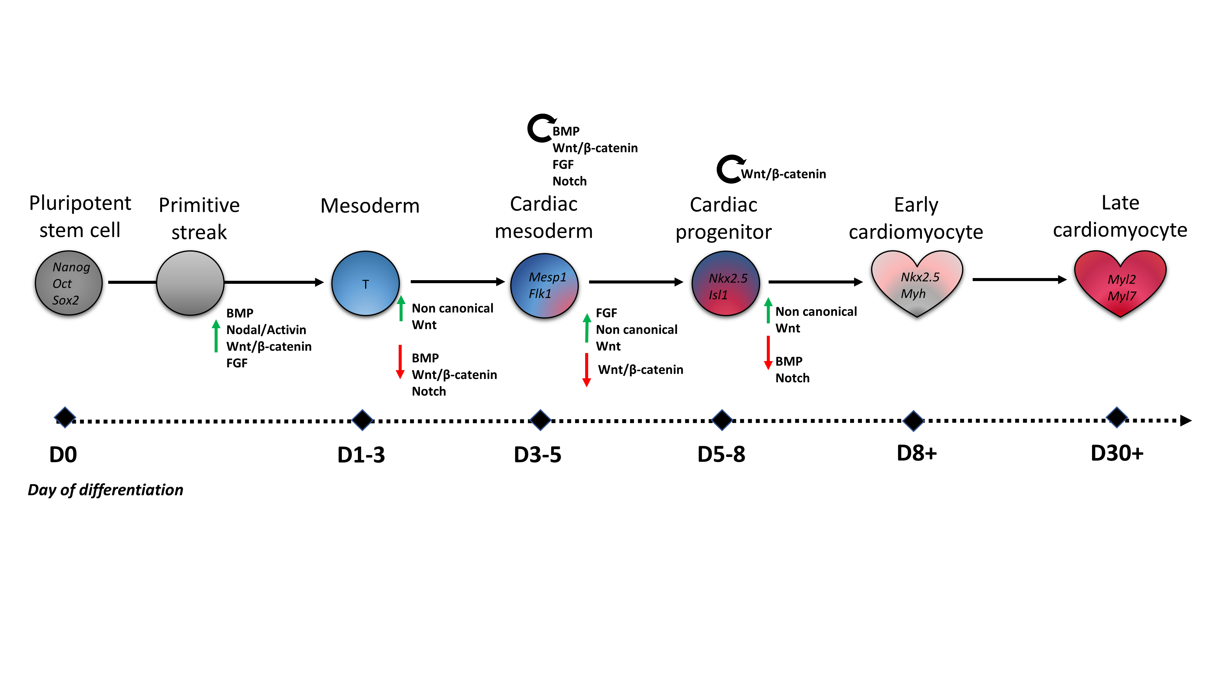
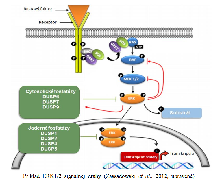
Mitogen activated protein kinases (MAPK) represent a family of proteins which is a part of MAPK signaling pathway and which is responsible for cells reaction to growth factors, hormones, stress, but also plays an important role in differentiation into many cell types. We have observed that there are changes in this signaling pathway in hypoxic conditions, then when certain chemical compounds are applied to cells as well as changes in phosphorylation of MAPK members during differentiation. MAPK are enzymatically active in their phosphorylated state and inactive in dephosphorylated. This inactivation can be carried out specifically by family of dual specificity phosphatases (DUSPs). Goal of one of our projects is shedding some light on what impact DUSP family has on MAPK pathway and subsequently on differentiation into cardiac cells.
If you are interested in our research and would like to learn more about it, or if you would like to colaborate in our research, do not hesitate to contact us. Information on why and how we study cardiomyogenesis can also be found final theses of our graduates.
Collaborations
- Doc. Ing. Petr Humpolíček, Ph.D. Tomas Bata University in Zlin, Faculty of Technology, Polymer Centre
- Doc. Mgr. Petr Vaňhara, Ph.D. Masaryk University, Faculty of Medicine
- Doc. MUDr. Markéta Bébarová, Ph.D. Masaryk University, Faculty of Medicine
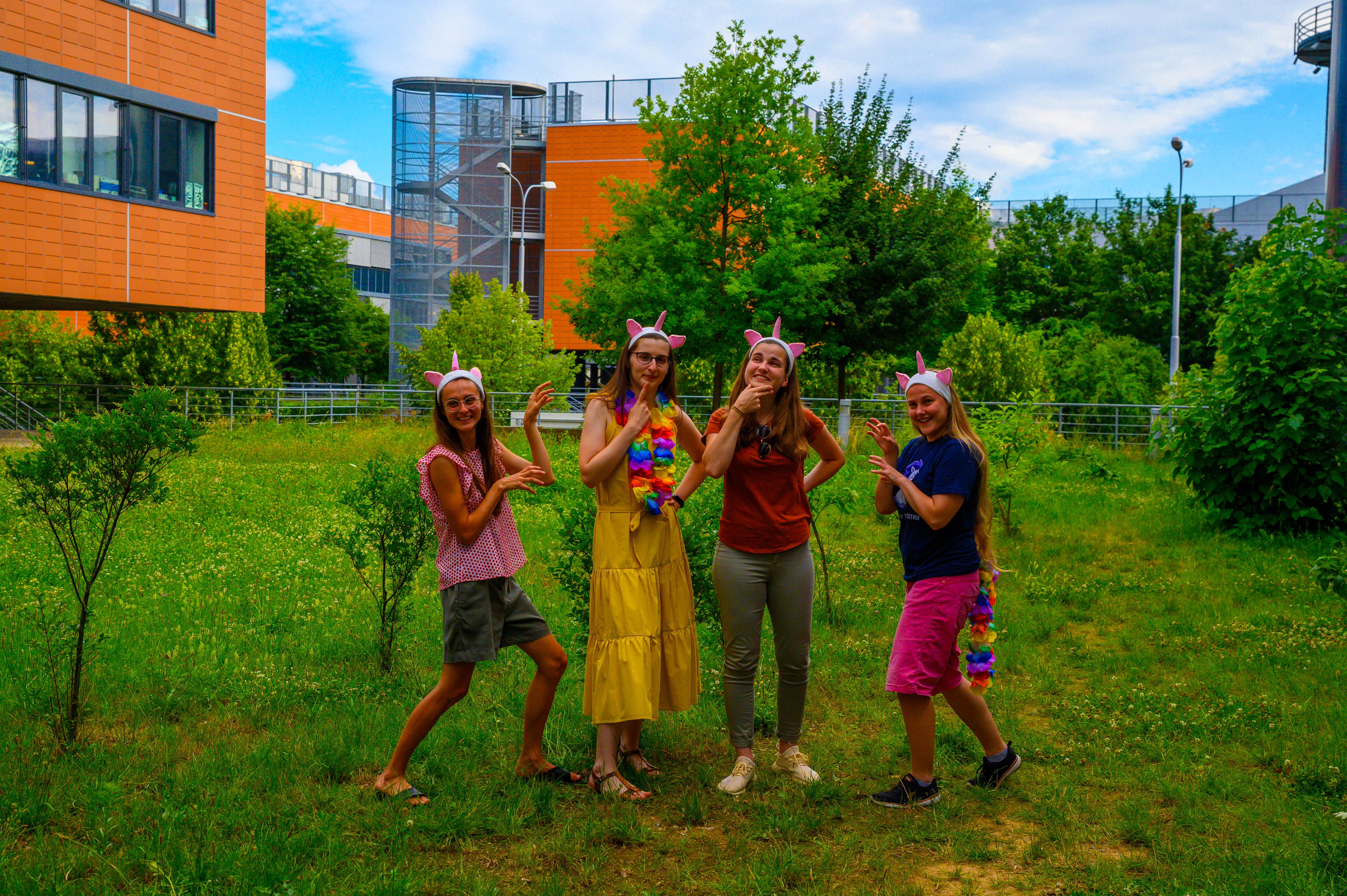
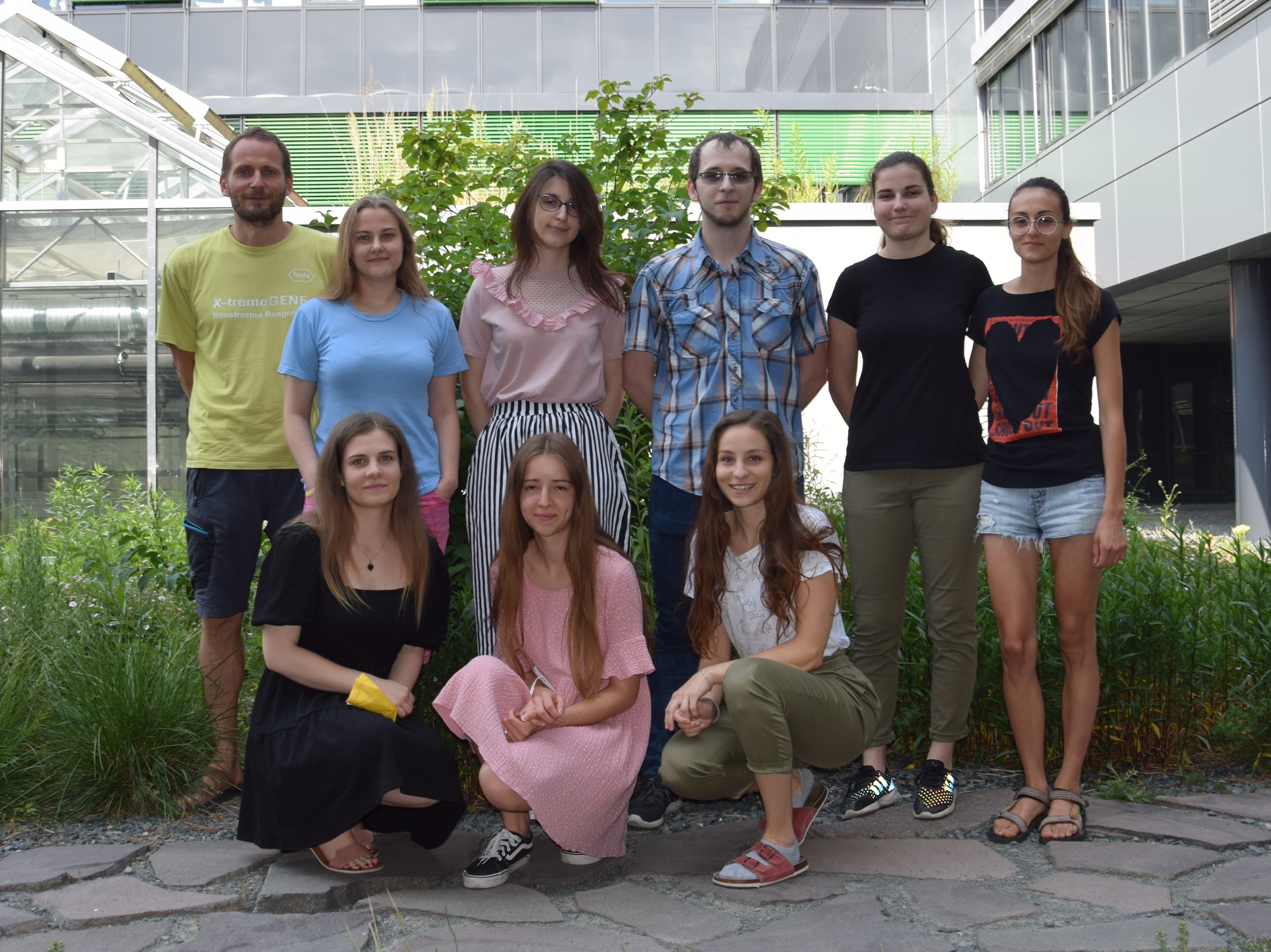
Research team:
Postdoc:
- Mgr. Stanislava Sladeček, Ph.D. (2022-)
- Katarzyna Anna Radaszkiewicz, Ph.D., M.Sc. (2017-2021)
- Josef Večeřa, Ph.D. (2012-2019)
Ph.D. students:
Alumni:
Mgr. students:
- Mgr. Zuzana Tomášiková
- Mgr. Martina Bőhmová
- Mgr. Markéta Mlčáková
- Mgr. Eliška Kohoutková
- Mgr. Katarína Streďanská
- Mgr. Petr Gintar
- Mgr. Simona Šmýkalová
- Mgr. Petra Lesáková
- Mgr. Bc. Deborah Beckerová
- Mgr. Lucie Woloszczuková
- Mgr. Veronika Šumberová
- Mgr. Vladislava Vodičková
- Mgr. Andrea Martinková
- Mgr. Vojtěch Koníř
- Mgr. Jana Moudrá
- Mgr. Veronika Pánská
- Mgr. Kateřina Kodymová
- Mgr. Bc. Radek Machát
- Mgr. Veronika Sikorová
- Mgr. Dominika Sýkorová
- Mgr. et Mgr. Martin Šodek
- Mgr. Marcela Kohoutková
- Mgr. Martin Kudláček
- Mgr. Markéta Hanáčková
- Mgr. Štěpánka Jankovská