Abstract:
Organ function depends on tissues adopting the correct architecture. However, insights into organ architecture are currently hampered by an absence of standardized quantitative 3D analysis.
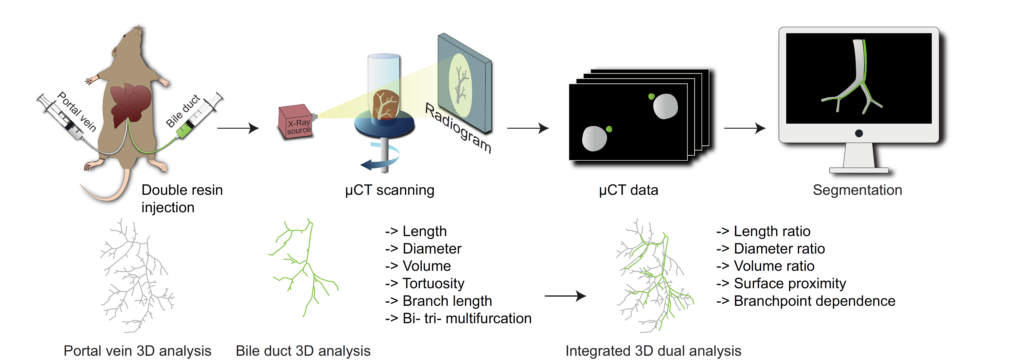
We aimed to develop a robust technology to visualize, digitalize, and segment the architecture of two tubular systems in 3D: double resin casting micro computed tomography (DUCT).
As proof of principle, we applied DUCT to a mouse model for Alagille syndrome (Jag1Ndr/Ndr mice), characterized by intrahepatic bile duct paucity, that can spontaneously generate a biliary system in adulthood. DUCT identified increased central biliary branching and peripheral bile duct tortuosity as two compensatory processes occurring in distinct regions of Jag1Ndr/Ndr liver, leading to full reconstitution of wild-type biliary volume and phenotypic recovery.
DUCT is thus a powerful new technology for 3D analysis, which can reveal novel phenotypes and provide a standardized method of defining liver architecture in mouse models.
Elife. 2021 Feb 26;10:e60916.doi: 10.7554/eLife.60916.
Authors:
Simona Hankeova #1,2, Jakub Salplachta #3, Tomas Zikmund #3, Michaela Kavkova #3, Noémi Van Hul #1, Adam Brinek 3, Veronika Smekalova 3, Jakub Laznovsky 3, Feven Dawit 4, Josef Jaros 5, Vítězslav Bryja 2, Urban Lendahl 6, Ewa Ellis 4, Antal Nemeth 7, Björn Fischler 4, Edouard Hannezo 8, Jozef Kaiser 3, Emma Rachel Andersson 1,6
- 1 Department of Biosciences and Nutrition, Karolinska Institutet, Solna, Sweden.
- 2 Department of Experimental Biology, Masaryk University, Brno, Czech Republic.
- 3 CEITEC – Central European Institute of Technology, Brno University of Technology, Brno, Czech Republic.
- 4 Department of Pediatrics, Clinical Science, Intervention and Technology (CLINTEC), Karolinska Institutet and Karolinska University Hospital, Solna, Sweden.
- 5 Department of Histology and Embryology, Masaryk University, Brno, Czech Republic.
- 6 Department of Cell and Molecular Biology, Karolinska Institutet, Solna, Sweden.
- 7 Department of Laboratory Medicine, Karolinska Institutet, Solna, Sweden.
- 8 Institute of Science and Technology, Klosterneuburg, Austria.
# Contributed equally.