Abstract
Organs throughout the body develop both asymmetrically and symmetrically. Here, we assess how symmetrical teeth in reptiles can be created from asymmetrical tooth germs.
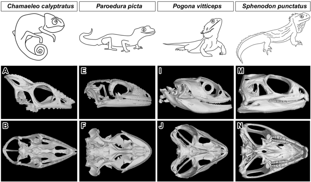
Teeth of lepidosaurian reptiles are mostly anchored to the jaw bones by pleurodont ankylosis, where the tooth is held in place on the labial side only. Pleurodont teeth are characterized by significantly asymmetrical development of the labial and lingual sides of the cervical loop, which later leads to uneven deposition of hard tissue. On the other hand, acrodont teeth found in lizards of the Acrodonta clade (i.e. agamas, chameleons) are symmetrically ankylosed to the jaw bone.
Here, we have focused on the formation of the symmetrical acrodont dentition of the veiled chameleon (Chamaeleo calyptratus). Intriguingly, our results revealed distinct asymmetries in morphology of the labial and lingual sides of the cervical loop during early developmental stages, both at the gross and ultrastructural level, with specific patterns of cell proliferation and stem cell marker expression. Asymmetrical expression of ST14 was also observed, with a positive domain on the lingual side of the cervical loop overlapping with the SOX2 domain. In contrast, micro-CT analysis of hard tissues revealed that deposition of dentin and enamel was largely symmetrical at the mineralization stage, highlighting the difference between cervical loop morphology during early development and differentiation of odontoblasts throughout later odontogenesis.
In conclusion, the early asymmetrical development of the enamel organ seems to be a plesiomorphic character for all squamate reptiles, while symmetrical and precisely orchestrated deposition of hard tissue during tooth formation in acrodont dentitions probably represents a novelty in the Acrodonta clade.
Authors
Kavková1,7, M. Šulcová2,6,7, J. Dumková3, O. Zahradníček4, J. Kaiser1, A. S. Tucker5,
Zikmund1 & M. Buchtová2,6
1Central European Institute of Technology, Brno University of Technology, Brno, Czech Republic
2Department of Experimental Biology, Faculty of Science, Masaryk University, Brno, Czech Republic
3Department of Histology and Embryology, Faculty of Medicine, Masaryk University, Brno, Czech Republic
4Department of Radiation Dosimetry, Nuclear Physics Institute, Czech Academy of Sciences, Prague, Czech Republic
5Centre for Craniofacial and Regenerative Biology, King’s College London, Floor 27 Guy’s Tower, Guy’s Hospital, London Bridge, London, UK
6Laboratory of Molecular Morphogenesis, Institute of Animal Physiology and Genetics, Czech Academy of Sciences, Veveri 97, 602 00 Brno, Czech Republic