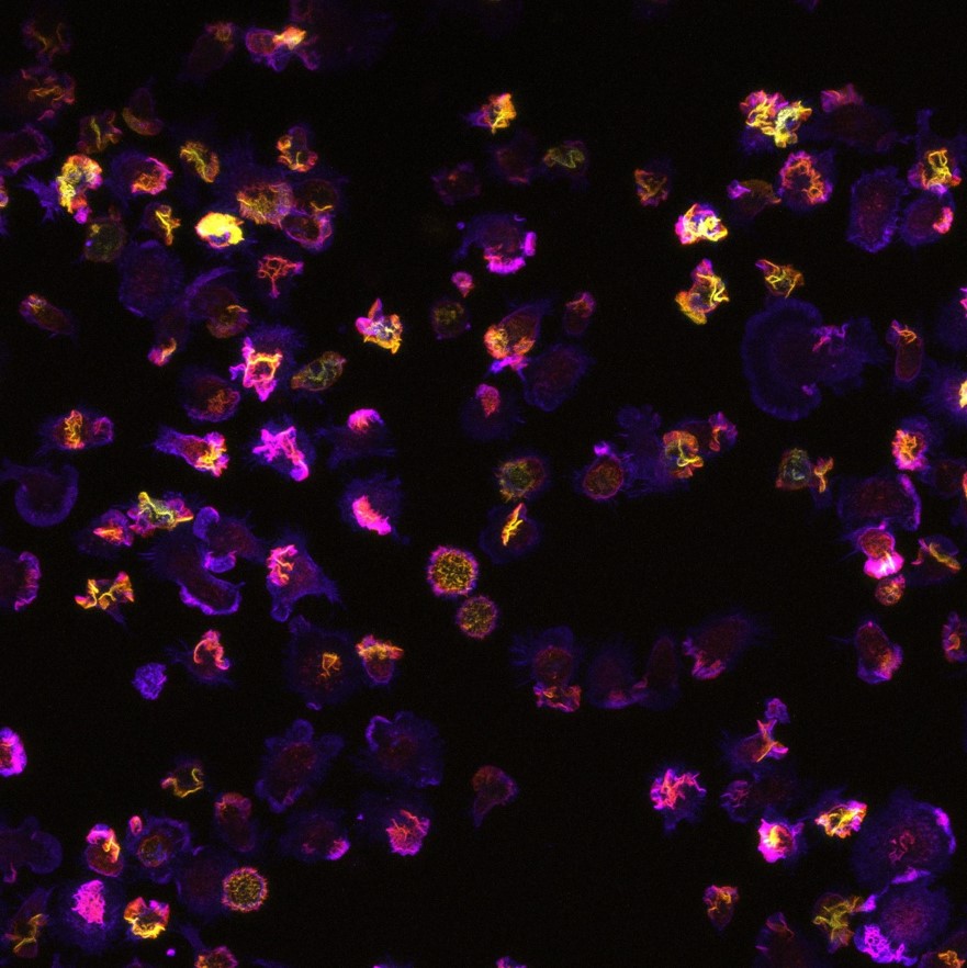
Thanks to the confocal microscope, we can observe the cells really closely! Take a look at a photograph of cells on which actin, a part of the cytoskeleton of cells, is highlighted with phalloidin.
The confocal microscope can capture the cell in multiple planes, and the software can then distinguish between the bottom and top of the cell. In yellow you can see the part of the cell closer to the lens of the microscope and the pink color marks the lower part of the cell.
The shape of the cells is not uniform, it is variable depending on what activity the cell is currently performing. For example, the longer shape of the cell indicates that the cell is in motion and is moving from place to place.

The author of the photo is Mgr. Štěpán Čada, whose PhD project deals with the role of non-canonical Wnt signaling in establishing polarity during amoboid cell migration.


